Here are the first images of how coronavirus replicates in cells. #Bloomberg
2227
Saturday, 01 February, 2020, 16:05
 Magnified pictures of the microbial cause of the pneumonia outbreak that’s sparked a global health emergency were released by scientists at the University of Hong Kong Friday. Thin-section electron micrographs of the novel coronavirus show part of an infected cell, grown in a culture, with virus particles being released from the cell’s surface. Each infected cell produces thousands of new infectious virus particles that can go on to infect new cells, said John Nicholls, a clinical professor of pathology at the university, who grew the virus with colleagues Leo Poon and Malik Peiris. 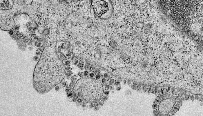 |

Healthy diet too expensive for one in three people globally, UN report finds
69922.07.2026, 00:52
Confirmed Ebola cases in Democratic Republic of Congo rise to 1,963
68114.07.2026, 16:34
Martha Lillard, last known US polio survivor using iron lung, dies aged 78 (photo)
74713.07.2026, 23:41
Uganda finds isolated Marburg virus case, Africa CDC says
150802.07.2026, 00:58
The WHO places suspicious Ebola cases at over 500 and deaths at 130
134119.05.2026, 18:13
Countries airlift nationals evacuated from virus-hit cruise ship
146010.05.2026, 23:47